おすすめの製品
由来生物
rabbit
品質水準
抗体製品の状態
purified immunoglobulin
抗体製品タイプ
primary antibodies
クローン
polyclonal
化学種の反応性
mouse, human, rat
メーカー/製品名
Upstate®
テクニック
immunohistochemistry: suitable
western blot: suitable
アイソタイプ
IgG
NCBIアクセッション番号
UniProtアクセッション番号
輸送温度
wet ice
ターゲットの翻訳後修飾
unmodified
遺伝子情報
human ... CASP3(836)
詳細
Caspase-3 (UniProt: P42574; also known as EC:3.4.22.56, CASP-3, Apopain, Cysteine protease CPP32, CPP-32, Protein Yama, SREBP cleavage activity 1, SCA-1) is encoded by the CASP3 (also known as CPP32) gene (Gene ID: 836) in human. Cysteine-aspartic proteases or Caspases play essential roles in apoptosis, necrosis, and inflammation. Historically, caspases were numbered in the order in which they were identified. Caspase-3 is a heterotetrameric enzyme that consists of two anti-parallel arranged heterodimers, each one formed by a 17 kDa (p17) and a 12 kDa (p12) subunit. Caspase-3 is initially produced with a propeptide sequence (aa 1-9), the removal of which yields the 268 aa. caspase-3 proenzyme. Upon activation, the proenzyme is proteolytically cleaved first between Asp175-Ser176 to generate a p20 (aa 10-175) fragment and the p12 (aa 176-277) subunit. Further cleavage of the p20 fragment between Asp28-Ser29 produces the p17 (aa 29-175) subunit. The p17 and p12 subunits dimerize and forms the active caspase-3 enzyme. Caspase-3 has a strict requirement for an Asp residue at positions P1 and P4. It has a preferred cleavage sequence of Asp-Xaa-Xaa-Asp-|- with a hydrophobic amino-acid residue at P2 and a hydrophilic amino-acid residue at P3, although Val or Ala are also accepted at this position. Caspase-3 is involved in the activation cascade of caspases responsible for apoptosis execution. At the onset of apoptosis, it proteolytically cleaves poly(ADP-ribose) polymerase (PARP) at a Asp216-|-Gly217 bond. Caspase-3 mediates the proteolytic activation of caspases-6 and -7, while caspase-3 itself is processed and activated by caspase-8, -9, or -10.
特異性
Recognizes full-length Caspase 3 (Yama/Apopain) and proteolytic fragments.
免疫原
Human full-length Caspase 3 fusion protein containing a histidine-6 tag
アプリケーション
This Anti-Caspase 3 Antibody is validated for use in Immunihistochmistry and Western Blotting for the detection of Caspase 3.
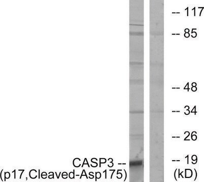
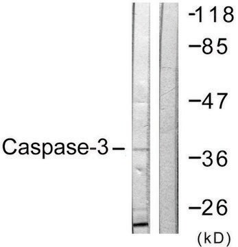
Western Blotting Analysis: 1μg/mL of this antibody detects Caspase-3 in A431 cell lysate.
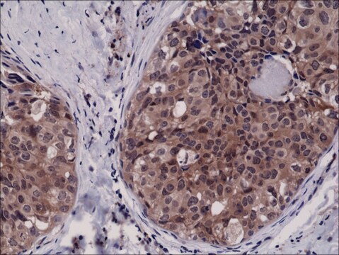
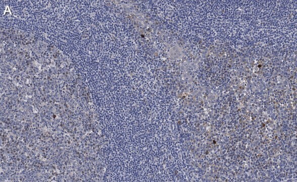
Immunohistochemistry (Paraffin) Analysis: A 1:250 dilution of this antibody detected Caspase-3 in Human tonsil tissue sections.
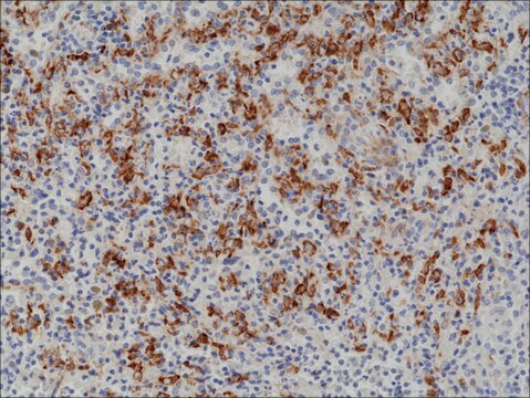
Immunohistochemistry (Paraffin) Analysis: A 1:250 dilution of this antibody detected Caspase-3 in Human tonsil tissue sections.
品質
routinely evaluated by immunoblot on RIPA lysates from non-stimulated human A431 cells, mouse 3T3/A31 or rat PC12 cells
ターゲットの説明
32 kDa
関連事項
Replaces: 04-1090; 04-439
物理的形状
Format: Purified
Protein A purified IgG in of 0.1M Tris-glycine, pH 7.4, 0.15M NaCl,and 0.05% sodium azide.
保管および安定性
Stable for 2 years at 2-8°C from date of shipment. For maximum recovery of product, centrifuge the original vial prior to removing the cap.
アナリシスノート
Control
Positive Antigen Control: Catalog #12-301, non-stimulated A431 cell lysate. Add 2.5µL of 2-mercaptoethanol/100µL of lysate and boil for 5 minutes to reduce the preparation. Load 20µg of reduced lysate per lane for mingels.
Positive Antigen Control: Catalog #12-301, non-stimulated A431 cell lysate. Add 2.5µL of 2-mercaptoethanol/100µL of lysate and boil for 5 minutes to reduce the preparation. Load 20µg of reduced lysate per lane for mingels.
法的情報
UPSTATE is a registered trademark of Merck KGaA, Darmstadt, Germany
適切な製品が見つかりませんか。
製品選択ツール.をお試しください
保管分類コード
10 - Combustible liquids
WGK
WGK 1
適用法令
試験研究用途を考慮した関連法令を主に挙げております。化学物質以外については、一部の情報のみ提供しています。 製品を安全かつ合法的に使用することは、使用者の義務です。最新情報により修正される場合があります。WEBの反映には時間を要することがあるため、適宜SDSをご参照ください。
Jan Code
06-735:
試験成績書(COA)
製品のロット番号・バッチ番号を入力して、試験成績書(COA) を検索できます。ロット番号・バッチ番号は、製品ラベルに「Lot」または「Batch」に続いて記載されています。
この製品を見ている人はこちらもチェック
M Leist et al.
Biochemical and biophysical research communications, 258(1), 215-221 (1999-05-01)
The endogenous mediator nitric oxide (NO) blocked apoptosis of Jurkat cells elicited by staurosporine, anti-CD95 or chemotherapeutics, and switched death to necrosis. The switch in the mode of cell death was dependent on the ATP loss elicited by NO. This
Antiandrogen-induced cell death in LNCaP human prostate cancer cells.
Lee, EC; Zhan, P; Schallhom, R; Packman, K; Tenniswood, M
Cell Death and Differentiation null
Prisca Boisguérin et al.
Cardiovascular research, 116(3), 633-644 (2019-05-31)
Regulated cell death is a main contributor of myocardial ischaemia-reperfusion (IR) injury during acute myocardial infarction. In this context, targeting apoptosis could be a potent therapeutical strategy. In a previous study, we showed that DAXX (death-associated protein) was essential for
A caspase cascade regulating developmental axon degeneration.
Simon, DJ; Weimer, RM; McLaughlin, T; Kallop, D; Stanger, K; Yang, J; O'Leary et al.
The Journal of Neuroscience null
Wafaa S Ramadan et al.
Cells, 10(12) (2021-12-25)
Patients suffering from Alzheimer's disease (AD) are still increasing worldwide. The development of (AD) is related to oxidative stress and genetic factors. This study investigated the therapeutic effects of ellagic acid (EA) on the entorhinal cortex (ERC), which plays a
ライフサイエンス、有機合成、材料科学、クロマトグラフィー、分析など、あらゆる分野の研究に経験のあるメンバーがおります。.
製品に関するお問い合わせはこちら(テクニカルサービス)