おすすめの製品
由来生物
mouse
品質水準
抗体製品の状態
purified antibody
抗体製品タイプ
primary antibodies
クローン
AE1/AE3, monoclonal
化学種の反応性
human
メーカー/製品名
Chemicon®
IHC Select
テクニック
immunohistochemistry: suitable (paraffin)
アイソタイプ
IgG1
輸送温度
wet ice
ターゲットの翻訳後修飾
unmodified
遺伝子情報
human ... KRT1(3848)
詳細
Keratins are a group of water-insoluble proteins that form monofilaments, a class of intermediate filament. These filaments form part of the cytoskeletal complex in epidermis and in most other epithelial tissues. Nineteen human epithelial keratins are resolved with two-dimensional gels electrophoresis (Moll, R., 1982). These can be divided into acid (pI <5.7) and basic (pI >6.0) subfamilies. The acidic keratins have molecular weights of 56.5, 55, 51, 50, 50′, 48, 46, 45, and 40 kD. The basic keratins have molecular weights of 65-67, 64, 59, 58, 56, and 52 kD. Members of the acidic and basic subfamilies are found together in pairs. The composition of keratin pairs varies with the epithelial cell type, stage of differentiation, cellular growth environment, and disease state (Sun, T.T., 1984; Cooper, D., 1985; Sun, T. T., 1985 ). The 56.5/65-67 kD pair is present in keratinized (differentiated) epidermis. The 55/64 kD pair is characteristic of normal (corneal-type) epithelial differentiation (Moll, R., 1982; Sun, T.T, 1984). The 51/59 kD pair is characteristic of the stratified squamous epithelial of internal organism such as esophagus and tongue (Moll, R., 1982; Cooper, D., 1985). The 51/58 kD pair is a keratinocyte marker; this pair is present in almost all stratified epithelia irrespective of the state of cellular stratification (Moll, R., 1982; Sun, T.T., 1984). The 48/56 kD pair is characteristic of hyper-proliferative (de-differentiated) keratinocytes (Moll, R., 1982; Weiss, R.A., 1984). The 45/52 kD pair and to a lesser extent, the 46/54 kD pair are characteristic of simple epithelia (Moll, R., 1982). The 40 kD keratin is present in most epithelia except adult epidermis (Moll, R., 1982).
特異性
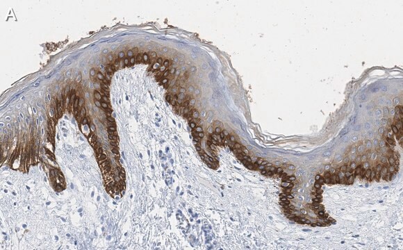
Anti-Keratin AE1 recognizes the 56.5, 50, 50′, 48, and 40 kD keratins of the acidic subfamily. Anti-keratin AE3 recognizes all members of the basic subfamily. AE1/AE3 reacts with both acidic and basic keratins. Staining is localized to the cytoplasm. AE1/AE3 specifically binds to antigens located in the cytoplasm of normal epithelia. Reacts with cells of epithelial origin including simple and stratified epithelia and epidermis.
免疫原
Human epidermal keratin
アプリケーション
Research Category
細胞骨格
細胞骨格
Research Sub Category
サイトケラチン
サイトケラチン
Antibody is prediluted and ready to use for Immunohistochemistry of formalin-fixed, paraffin-embedded tissues.
Pretreatment: Heat Induced Epitope Retrieval (HIER). Recommend Citrate Buffer, pH 6.0 (Cat. No. 21545). No signal was detected without Epitope retrieval.
Incubation: 10 minutes with IHC Select Detection Kits.
Cytokeratin AE1/AE3 has been prediluted for use as the primary antibody with Chemicon′s IHC Select Detection Kits and Protocols (Catalog Nos. DAB050, DET-HP1000, APR050, and DET-APR1000), but other supplier′s IHC detection systems may be used. For optimized protocol details, visit www.chemicon.com and select the protocols link under Cat. No.IHCR2025-6.
IHC Select Anti-Cytokeratin AE1/AE3 (Pan cytokeratins) Antibody, prediluted, clone AE1/AE3 is an antibody against Cytokeratin AE1/AE3 (Pan cytokeratins) for use in IH(P).
物理的形状
Format: Purified
Purified monoclonal mouse antibody supplied in liquid format diluted in PBS, pH 7.2 with stabilizers, 0.2% Tween 20, and 0.1% Kathon as preservative.
保管および安定性
Maintain at 2-8°C . Refer to vial for expiration dating.
法的情報
CHEMICON is a registered trademark of Merck KGaA, Darmstadt, Germany
免責事項
Unless otherwise stated in our catalog or other company documentation accompanying the product(s), our products are intended for research use only and are not to be used for any other purpose, which includes but is not limited to, unauthorized commercial uses, in vitro diagnostic uses, ex vivo or in vivo therapeutic uses or any type of consumption or application to humans or animals.
適切な製品が見つかりませんか。
製品選択ツール.をお試しください
シグナルワード
Warning
危険有害性情報
危険有害性の分類
Aquatic Chronic 3 - Skin Sens. 1
保管分類コード
12 - Non Combustible Liquids
WGK
WGK 2
引火点(°F)
Not applicable
引火点(℃)
Not applicable
適用法令
試験研究用途を考慮した関連法令を主に挙げております。化学物質以外については、一部の情報のみ提供しています。 製品を安全かつ合法的に使用することは、使用者の義務です。最新情報により修正される場合があります。WEBの反映には時間を要することがあるため、適宜SDSをご参照ください。
Jan Code
IHCR2025-6:
試験成績書(COA)
製品のロット番号・バッチ番号を入力して、試験成績書(COA) を検索できます。ロット番号・バッチ番号は、製品ラベルに「Lot」または「Batch」に続いて記載されています。
Cindy Perez-Pacheco et al.
Clinical cancer research : an official journal of the American Association for Cancer Research, 29(13), 2501-2512 (2023-04-12)
Perineural invasion (PNI) in oral cavity squamous cell carcinoma (OSCC) is associated with poor survival. Because of the risk of recurrence, patients with PNI receive additional therapies after surgical resection. Mechanistic studies have shown that nerves in the tumor microenvironment
Ligia B Schmitd et al.
Neoplasia (New York, N.Y.), 20(7), 657-667 (2018-05-26)
A diagnosis of perineural invasion (PNI), defined as cancer within or surrounding at least 33% of the nerve, leads to selection of aggressive treatment in squamous cell carcinoma (SCC). Recent mechanistic studies show that cancer and nerves interact prior to
Jing Kong et al.
Cell communication & adhesion, 24(1), 11-18 (2018-05-08)
Salivary gland adenoid cystic carcinoma (SACC) is one of the most common malignancies in the oral and maxillofacial region. Carcinoma-associated fibroblast (CAF) is an important component in the tumor microenvironment and participates in SACC progression. In this study, we established
Christina Springstead Scanlon et al.
Nature communications, 6, 6885-6885 (2015-04-29)
Perineural invasion (PNI) is an indicator of poor survival in multiple cancers. Unfortunately, there is no targeted treatment for PNI since the molecular mechanisms are largely unknown. PNI is an active process, suggesting that cancer cells communicate with nerves. However
ライフサイエンス、有機合成、材料科学、クロマトグラフィー、分析など、あらゆる分野の研究に経験のあるメンバーがおります。.
製品に関するお問い合わせはこちら(テクニカルサービス)


