MAB326
Anti-CNPase Antibody, clone 11-5B
clone 11-5B, Chemicon®, from mouse
Synonym(s):
2′3′-Cyclic Nucleotide 3′-Phosphohydrolase
About This Item
Recommended Products
biological source
mouse
Quality Level
antibody form
purified immunoglobulin
antibody product type
primary antibodies
clone
11-5B, monoclonal
species reactivity
canine, sheep, mouse, pig, bovine, rat, rabbit, human
should not react with
guinea pig, chicken
manufacturer/tradename
Chemicon®
technique(s)
immunocytochemistry: suitable
immunohistochemistry (formalin-fixed, paraffin-embedded sections): suitable
western blot: suitable
isotype
IgG1
NCBI accession no.
UniProt accession no.
shipped in
wet ice
target post-translational modification
unmodified
Gene Information
human ... CNP(1267)
General description
Immunohistochemical localization of CNPase has shown the enzyme to be restricted to oligodendrocytes and Schwann cells. The enzyme appears to be distributed in single and double loose wraps of myelin and not in compact myelin as earlier thought by most investigators. CNPase is located on the inner and outer loops of myelin, paranodally and near the inner surface of the oligodendrocyte membrane. In plaque regions of multiple sclerosis patients, the enzyme is reduced, and when CNS myelin is decreased, CNPase is one of the earlier proteins to be lost from the myelin. In addition, an enzyme that is probably identical to brain CNPase is located in the outer rod segments within the visual system, and this protein is also recognized by the monoclonal antibody 11-5B. In mixed human gliomas, the enzyme levels are reduced, although about 5% of the oligodendrocytes occasionally show normal positive staining.
Specificity
Immunogen
Application
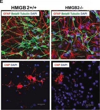
A previous lot was used on primary oligodendrocyte cultures.
Immunohistochemistry:
A previous lot was used on rat hippocampus tissue and rat spinal cord.
Immunoblotting of myelin, the Wolfgram protein fraction, the SN4 fraction, tissue sections and mixed glial tumors (oligodendrogliomas, etc.) CNPase I (46 kDa) and CNPase II (48 kDa), which are differentially regulated during development, with the larger protein being expressed earlier than CNPase I during development.
Immunohistochemistry on both fresh frozen and paraffin embedded tissue (microwave pretreatment, ctirate pH 6.0).
Immunoblot:
Immunoblotting of myelin, the Wolfgram protein fraction, the SN4 fraction, tissue sections
and mixed glial tumors (oligodendrogliomas, etc.)
Optimal working dilutions must be determined by end user.
Neuroscience
Neuronal & Glial Markers
Quality
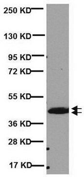
Western Blot Analysis:
1:1000 dilution of this lot detected CNPASE on 10 μg of Mouse Brain lysates.
Target description
Physical form
Storage and Stability
Analysis Note
Western Blot: Fresh bovine whole brain extract, mouse brain lysate.
Immunohistochemistry: Rat hippocampus tissue, rat spinal cord tissue.
Other Notes
Legal Information
Disclaimer
Not finding the right product?
Try our Product Selector Tool.
recommended
Storage Class
10 - Combustible liquids
wgk_germany
WGK 2
Certificates of Analysis (COA)
Search for Certificates of Analysis (COA) by entering the products Lot/Batch Number. Lot and Batch Numbers can be found on a product’s label following the words ‘Lot’ or ‘Batch’.
Already Own This Product?
Find documentation for the products that you have recently purchased in the Document Library.
Our team of scientists has experience in all areas of research including Life Science, Material Science, Chemical Synthesis, Chromatography, Analytical and many others.
Contact Technical Service


