Sign In to View Organizational & Contract Pricing.
Select a Size
Change View
About This Item
Conjugate:
unconjugated
Clone:
polyclonal
Application:
IHC
Citations:
13
biological source
rabbit
Quality Level
conjugate
unconjugated
antibody form
affinity isolated antibody
antibody product type
primary antibodies
clone
polyclonal
product line
Prestige Antibodies® Powered by Atlas Antibodies
form
buffered aqueous glycerol solution
species reactivity
human
technique(s)
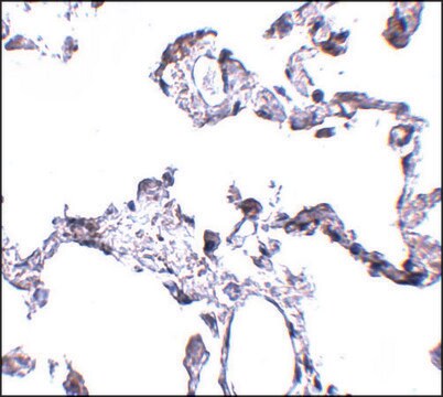
immunohistochemistry: 1:500-1:1000
immunogen sequence
RDKVMSEFNNNFRQQMENYPKNNHTASILDRMQADFKCCGAANYTDWEKIPSMSKNRVPDSCCINVTVGCGINFNEKAIHKEGCVEKIG
UniProt accession no.
application(s)
research pathology
shipped in
wet ice
storage temp.
−20°C
target post-translational modification
unmodified
Gene Information
human ... CD63(967)
General description
CD63 (cluster of differentiation) is a membrane protein belonging to the tetraspanin family of proteins called LAMP (lysosome-associated membrane glycoprotein) proteins. It is localized to histamine-containing granules.
It also resides in MHC (major histocompatibility compartment) II endocytic compartment.
Immunogen
CD63 antigen recombinant protein epitope signature tag (PrEST)
Application
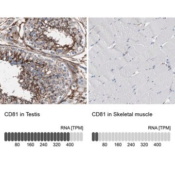
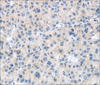
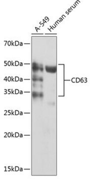
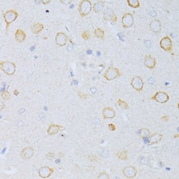
All Prestige Antibodies Powered by Atlas Antibodies are developed and validated by the Human Protein Atlas (HPA) project and as a result, are supported by the most extensive characterization in the industry.
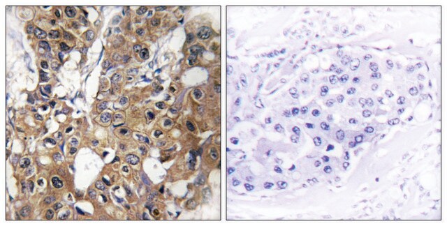
The Human Protein Atlas project can be subdivided into three efforts: Human Tissue Atlas, Cancer Atlas, and Human Cell Atlas. The antibodies that have been generated in support of the Tissue and Cancer Atlas projects have been tested by immunohistochemistry against hundreds of normal and disease tissues and through the recent efforts of the Human Cell Atlas project, many have been characterized by immunofluorescence to map the human proteome not only at the tissue level but now at the subcellular level. These images and the collection of this vast data set can be viewed on the Human Protein Atlas (HPA) site by clicking on the Image Gallery link. We also provide Prestige Antibodies® protocols and other useful information.
Biochem/physiol Actions
CD63 (cluster of differentiation) is associated with neutrophil homeostasis in a TIMP-1 (tissue inhibitor of metalloproteinases-1)-mediated manner. This protein is also involved in promoting metastasis, through the stabilization of β-catenin. It also affects the plasticity of cell. Its expression is high during early stages of melanoma, which decreases with the progression of melanoma. Thus, it functions as a tumor suppressor by inhibiting EMT (epithelial-mesenchymal transition) in melanoma. CD63, along with CD9, prevents cell-cell fusion in HIV (human immunodeficiency virus) infection, during transition from hemifusion to pore opening.
Features and Benefits
Prestige Antibodies® are highly characterized and extensively validated antibodies with the added benefit of all available characterization data for each target being accessible via the Human Protein Atlas portal linked just below the product name at the top of this page. The uniqueness and low cross-reactivity of the Prestige Antibodies® to other proteins are due to a thorough selection of antigen regions, affinity purification, and stringent selection. Prestige antigen controls are available for every corresponding Prestige Antibody and can be found in the linkage section.
Every Prestige Antibody is tested in the following ways:
- IHC tissue array of 44 normal human tissues and 20 of the most common cancer type tissues.
- Protein array of 364 human recombinant protein fragments.
Physical form
Solution in phosphate-buffered saline, pH 7.2, containing 40% glycerol and 0.02% sodium azide
Other Notes
Corresponding Antigen APREST72477
Legal Information
Prestige Antibodies is a registered trademark of Merck KGaA, Darmstadt, Germany
Disclaimer
Unless otherwise stated in our catalog or other company documentation accompanying the product(s), our products are intended for research use only and are not to be used for any other purpose, which includes but is not limited to, unauthorized commercial uses, in vitro diagnostic uses, ex vivo or in vivo therapeutic uses or any type of consumption or application to humans or animals.
Still not finding the right product?
Explore all of our products under Anti-CD63 antibody produced in rabbit
— or —
Try our Product Selector Tool to narrow your options
Storage Class
10 - Combustible liquids
wgk
WGK 1
flash_point_f
Not applicable
flash_point_c
Not applicable
ppe
Eyeshields, Gloves, multi-purpose combination respirator cartridge (US)
Choose from one of the most recent versions:
Already Own This Product?
Find documentation for the products that you have recently purchased in the Document Library.
Shin La Shu et al.
Methods in molecular biology (Clifton, N.J.), 2265, 289-304 (2021-03-12)
Cells release extracellular vesicles (EVs) that can be detected both in vivo and in cell culture medium. Among EVs, exosomes are 50-150 nm vesicles that are systematically packaged into multivesicular bodies for release into the external environment. In cancer, these intentionally
Sandra Pinet et al.
Oncotarget, 7(31), 50349-50364 (2016-07-08)
The neurotrophin receptors are known to promote growth and proliferation of glioblastoma cells. Their functions in spreading glioblastoma cell aggressiveness to the microenvironment through exosome release from glioblastoma cells are unknown. Considering previous reports demonstrating that YKL-40 expression is associated
Julia Kobuch et al.
Haematologica, 100(8), 1005-1013 (2015-05-24)
The homeostasis of neutrophil granulocytes can affect the outcome of several inflammation-associated diseases including cancer. The regulation of this homeostasis is still not completely understood. We previously found that elevated systemic levels of tissue inhibitor of metalloproteinases-1 (TIMP-1) induce an
Global Trade Item Number
| SKU | GTIN |
|---|---|
| HPA010088-100UL | 04061837125218 |
| HPA010088-25UL | 04061842816354 |