Skip to Content
MilliporeSigma
All Photos(2)

Documents

SAB4200813

Sigma-Aldrich

Anti-α Actinin antibody, Mouse monoclonal

clone BM-75.2, purified from hybridoma cell culture

Synonym(s):

Alpha-actinin cytoskeletal isoform, Alpha-actinin-1, F-actin cross-linking protein, Non-muscle alpha-actinin-1

Sign Into View Organizational & Contract Pricing


About This Item

UNSPSC Code:
12352203
NACRES:
NA.41

biological source

mouse

antibody form

purified from hybridoma cell culture

antibody product type

primary antibodies

clone

BM-75.2, monoclonal

mol wt

~100 kDa

species reactivity

human

packaging

antibody small pack of 25 μL

concentration

~1 mg/mL

technique(s)

immunoblotting: 0.5-1 μg/mL using human HeLa cells extract.
immunofluorescence: 5-10 μg/mL using human foreskin fibroblast Hs68 cells.

isotype

IgM

UniProt accession no.

shipped in

dry ice

Storage temp.

−20°C

target post-translational modification

unmodified

Gene Information

human ... ACTN1(87)

General description

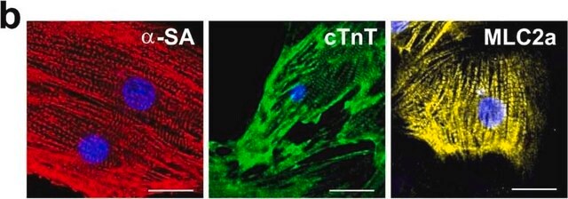
Monoclonal Anti- α Actinin (mouse IgM isotype) is derived from the hybridoma BM-72.5 produced by the fusion of mouse myeloma cells and splenocytes from mice immunized with a cytoskeletal fraction of bovine mammary gland epithelium (BMGE) cultured cells. α-Actinin is an actin binding protein present in both muscle and non-muscle cells. It has a molecular weight of 100,000 daltons and forms dimers in solution. In normal skeletal muscle, α-actinin is associated with the z-discs that define muscle sarcomeres. In smooth muscle, α-actinin has been detected in dense bodies and plaques characteristic of the tissue.

Specificity

Monoclonal Anti-α-Actinin specifically recognizes α-Actinin from human1, mouse2, rat3, chicken4, monkey, canine and bovine origin. 

Immunogen

Cytoskeletal fraction of bovine mammary gland epithelium (BMGE) cultured cells.

Application

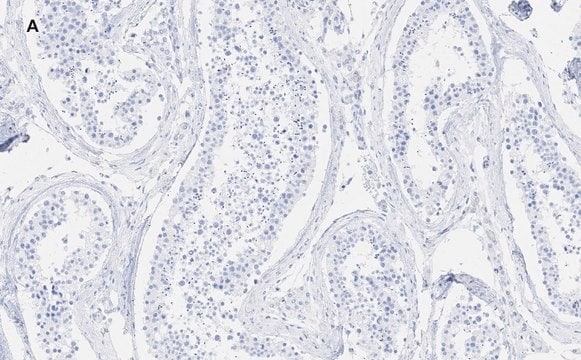
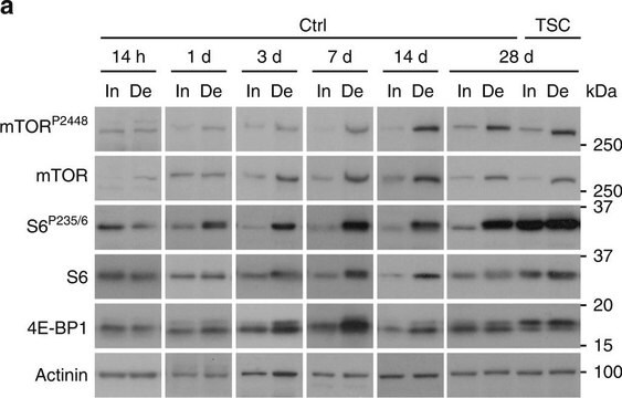
Anti-α Actinin antibody may be used in various immunochemical techniques including Immunoblot (~100 kDa), Immunohistochemistry and Immunofluorescence. Monoclonal Anti-α-Actinin may be used for immunofluorescent localization of α-actinin in cultured cells and tissues, for immunofluorescent labeling of skeletal muscle (normal and pathological) in order to detect its organizational state, studies of membrane anchorage sites and immunochemical identification of α-actinin by immunoblotting analysis.

Biochem/physiol Actions

α-actinin has extensive proteins association with actin containing stress fibers, in particular with their membrane bound termini.
α-actinin modulates muscle contraction and has a role in cytoskeletal organisation. α-actinin acts as an actin crosslinker and scaffold. Mutations in this gene has been linked to heterogeneous hypertrophic cardiomyopathy and juvenile onset atrial fibrillation.

Physical form

Supplied as a solution in 0.01 M phosphate buffered saline pH 7.4, containing 15 mM sodium azide as a preservative.

Storage and Stability

For continuous use, store at 2-8°C for up to one month. For extended storage, freeze in working aliquots. Repeated freezing and thawing is not recommended. If slight turbidity occurs upon prolonged storage, clarify the solution by centrifugation before use. Working dilution samples should be discarded if not used within 12 hours.

Disclaimer

Unless otherwise stated in our catalog  our products are intended for research use only and are not to be used for any other purpose, which includes but is not limited to, unauthorized commercial uses, in vitro diagnostic uses, ex vivo or in vivo therapeutic uses or any type of consumption or application to humans or animals.

wgk_germany

nwg

flash_point_f

Not applicable

flash_point_c

Not applicable


Certificates of Analysis (COA)

Search for Certificates of Analysis (COA) by entering the products Lot/Batch Number. Lot and Batch Numbers can be found on a product’s label following the words ‘Lot’ or ‘Batch’.

Already Own This Product?

Find documentation for the products that you have recently purchased in the Document Library.

Visit the Document Library

Feng Zhang et al.
Materials today. Bio, 20, 100626-100626 (2023-05-01)
Heart-on-chip emerged as a potential tool for cardiac tissue engineering, recapitulating key physiological cues in cardiac pathophysiology. Controlled electrical stimulation and the ability to provide directly analyzed functional readouts are essential to evaluate the physiology of cardiac tissues in the
Novel alpha-actinin 2 variant associated with familial hypertrophic cardiomyopathy and juvenile atrial arrhythmias: a massively parallel sequencing study
Girolami F, et al.
Circulation. Cardiovascular Genetics, 7(6), 741-750 (2014)
Activation of human neutrophils induces an interaction between the integrin beta 2-subunit (CD18) and the actin binding protein alpha-actinin.
Pavalko FM and LaRoche SM
Journal of immunology (Baltimore, Md. : 1950), 151(7), 3795-3807 (1993)
The vertebrate muscle Z-disc: sarcomere anchor for structure and signalling
Luther PK
Journal of Muscle Research and Cell Motility, 30(5-6), 171-185 (2009)
Samaneh Yazdani et al.
Molecular biology of the cell, 33(12), ar106-ar106 (2022-08-04)
Endothelia determine blood-to-tissue solute delivery, yet glucose transit is poorly understood. To illuminate mechanisms, we tracked [3H]-2-deoxyglucose (2-DG) in human adipose-tissue microvascular endothelial cells. 2-DG uptake was largely facilitated by the glucose transporters GLUT1 and GLUT3. Once in the cytosol

Our team of scientists has experience in all areas of research including Life Science, Material Science, Chemical Synthesis, Chromatography, Analytical and many others.

Contact Technical Service