おすすめの製品
由来生物
rabbit
品質水準
抗体製品の状態
serum
抗体製品タイプ
primary antibodies
クローン
polyclonal
化学種の反応性
yeast, Saccharomyces cerevisiae, human
化学種の反応性(ホモロジーによる予測)
vertebrates (most common)
メーカー/製品名
Upstate®
テクニック
ChIP: suitable (ChIP-seq)
dot blot: suitable
western blot: suitable
NCBIアクセッション番号
UniProtアクセッション番号
輸送温度
dry ice
ターゲットの翻訳後修飾
acetylation (Lys18)
遺伝子情報
human ... H3C1(8350)
詳細
Histone H3 is one of the 5 main histone proteins involved in the structure of chromatin in eukaryotic cells. Featuring a main globular domain and a long N terminal tail H3 is involved with the structure of the nucleosomes of the ′beads on a string′ structure.
Acetylation of histone H3 occurs at several different lysine positions in the histone tail and is performed by a family of enzymes known as Histone Acetyl Transferases (HATs).
Acetylation of histone H3 occurs at several different lysine positions in the histone tail and is performed by a family of enzymes known as Histone Acetyl Transferases (HATs).
特異性
Recognizes Histone H3 acetylated on lysine 18.
免疫原
KLH-conjugated, synthetic peptide (GKAPRAcKQLASK-C) corresponding to amino acids 13-23 of yeast Histone H3 acetylated on lysine 18with a C-terminal cysteine added for conjugation purposes
アプリケーション
Chromatin Immunoprecipitation:
Representative lot data.
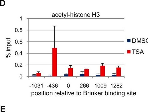
Sonicated chromatin prepared from HeLa cells (1 X 10E6 cell equivalents per IP) were subjected to chromatin immunoprecipitation using 2 µL of either Normal Rabbit Serum , or 2 µL of Anti-Acetyl-Histone H3 (Lys18)and the Magna ChIP A Kit (Cat. # 17-610). Successful immunoprecipitation of Acetyl-Histone H3 (Lys18) associated DNA fragments was verified by qPCR using Control Primers specific for the human GAPDH promoter region as a positive locus, and β-globin primers as a negative locus. Data is presented as percent input of each IP sample relative to input chromatin for each amplicon and ChIP sample as indicated.
Please refer to the EZ-Magna ChIP A (Cat. # 17-408) or EZ-ChIP (Cat. # 17-371) protocol for experimental details.
ChIP-Sequencing:
Representative lot data. Chromatin immunoprecipitation was performed using the Magna ChIP HiSens kit (cat# 17-10460), 2 μg of anti-acetyl-Histone H3 (Lys18) antibody (cat# 07-354), 20 µL Protein A/G beads , and 1e6 crosslinked HeLa cell chromatin followed by DNA purification using magnetic beads. Libraries were prepared from Input and ChIP DNA samples using standard protocols with Illumina barcoded adapters, and analyzed on Illumina HiSeq instrument. An excess of ten million reads from FastQ files were mapped using Bowtie (http://bowtie-bio.sourceforge.net/manual.shtml) following TagDust (http://genome.gsc.riken.jp/osc/english/dataresource/) tag removal. Peaks were identified using MACS (http://luelab.dfci.harvard.edu/MACS/), with peaks and reads visualized as a custom track in UCSC Genome Browser (http://genome.ucsc.edu) from BigWig and BED files.
Western Blot Analysis:
Representative lot data.
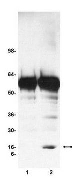
Recombinant histone H3 (lane 1, Catalog # 14-494) and acid extracts from sodium butyrate treated (lane 2) and untreated (lane 3) HeLa cells (Catalog # 17-305) were probed with anti acetyl- Histone H3 (Lys18) (1:10,000 dilution).
Arrow indicates acetyl histone H3 (Lys18) (17 kDa).
Dot Blot:
Representative lot data.
40 ng and 4ng amounts of histone peptides with various modifications (see table 1) were transferred to PVDF membrane and probed with Anti-Acetyl-Histone H3 (Lys18) antibody (1:5000 dilution). Proteins were visualized using a goat anti-rabbit IgG conjugated to HRP and a chemiluminescence detection system. Image from a 60 second exposure is shown.
Representative lot data.
Sonicated chromatin prepared from HeLa cells (1 X 10E6 cell equivalents per IP) were subjected to chromatin immunoprecipitation using 2 µL of either Normal Rabbit Serum , or 2 µL of Anti-Acetyl-Histone H3 (Lys18)and the Magna ChIP A Kit (Cat. # 17-610). Successful immunoprecipitation of Acetyl-Histone H3 (Lys18) associated DNA fragments was verified by qPCR using Control Primers specific for the human GAPDH promoter region as a positive locus, and β-globin primers as a negative locus. Data is presented as percent input of each IP sample relative to input chromatin for each amplicon and ChIP sample as indicated.
Please refer to the EZ-Magna ChIP A (Cat. # 17-408) or EZ-ChIP (Cat. # 17-371) protocol for experimental details.
ChIP-Sequencing:
Representative lot data. Chromatin immunoprecipitation was performed using the Magna ChIP HiSens kit (cat# 17-10460), 2 μg of anti-acetyl-Histone H3 (Lys18) antibody (cat# 07-354), 20 µL Protein A/G beads , and 1e6 crosslinked HeLa cell chromatin followed by DNA purification using magnetic beads. Libraries were prepared from Input and ChIP DNA samples using standard protocols with Illumina barcoded adapters, and analyzed on Illumina HiSeq instrument. An excess of ten million reads from FastQ files were mapped using Bowtie (http://bowtie-bio.sourceforge.net/manual.shtml) following TagDust (http://genome.gsc.riken.jp/osc/english/dataresource/) tag removal. Peaks were identified using MACS (http://luelab.dfci.harvard.edu/MACS/), with peaks and reads visualized as a custom track in UCSC Genome Browser (http://genome.ucsc.edu) from BigWig and BED files.
Western Blot Analysis:
Representative lot data.
Recombinant histone H3 (lane 1, Catalog # 14-494) and acid extracts from sodium butyrate treated (lane 2) and untreated (lane 3) HeLa cells (Catalog # 17-305) were probed with anti acetyl- Histone H3 (Lys18) (1:10,000 dilution).
Arrow indicates acetyl histone H3 (Lys18) (17 kDa).
Dot Blot:
Representative lot data.
40 ng and 4ng amounts of histone peptides with various modifications (see table 1) were transferred to PVDF membrane and probed with Anti-Acetyl-Histone H3 (Lys18) antibody (1:5000 dilution). Proteins were visualized using a goat anti-rabbit IgG conjugated to HRP and a chemiluminescence detection system. Image from a 60 second exposure is shown.
Use Anti-acetyl-Histone H3 (Lys18) Antibody (Rabbit Polyclonal Antibody) validated in ChIP, WB to detect acetyl-Histone H3 (Lys18) also known as H3K18Ac, Histone H3 (acetyl K18).
品質
routinely evaluated by immunoblot on acid extracts from sodium butyrate treated HeLa cells
ターゲットの説明
17 kDa
関連事項
Replaces: 04-1107
物理的形状
100 μLof rabbit serum containing 0.05% sodium azide and 30% glycerol.
その他情報
Concentration: Please refer to the Certificate of Analysis for the lot-specific concentration.
法的情報
UPSTATE is a registered trademark of Merck KGaA, Darmstadt, Germany
適切な製品が見つかりませんか。
製品選択ツール.をお試しください
保管分類コード
12 - Non Combustible Liquids
WGK
WGK 1
引火点(°F)
Not applicable
引火点(℃)
Not applicable
適用法令
試験研究用途を考慮した関連法令を主に挙げております。化学物質以外については、一部の情報のみ提供しています。 製品を安全かつ合法的に使用することは、使用者の義務です。最新情報により修正される場合があります。WEBの反映には時間を要することがあるため、適宜SDSをご参照ください。
Jan Code
07-354:
試験成績書(COA)
製品のロット番号・バッチ番号を入力して、試験成績書(COA) を検索できます。ロット番号・バッチ番号は、製品ラベルに「Lot」または「Batch」に続いて記載されています。
Inducible activation of Cre recombinase in adult mice causes gastric epithelial atrophy, metaplasia and regenerative changes in the absence of "floxed" alleles.
Huh WJ, Mysorekar IU, Mills JC
American Journal of Physiology: Gastrointestinal and Liver Physiology null
Paola Y Bertucci et al.
Nucleic acids research, 41(12), 6072-6086 (2013-05-04)
Steroid receptors were classically described for regulating transcription by binding to target gene promoters. However, genome-wide studies reveal that steroid receptors-binding sites are mainly located at intragenic regions. To determine the role of these sites, we examined the effect of
B Ruthrotha Selvi et al.
The Journal of biological chemistry, 285(10), 7143-7152 (2009-12-22)
Methylation of the arginine residues of histones by methyltransferases has important consequences for chromatin structure and gene regulation; however, the molecular mechanism(s) of methyltransferase regulation is still unclear, as is the biological significance of methylation at particular arginine residues. Here
Nucleosome competition reveals processive acetylation by the SAGA HAT module.
Ringel, AE; Cieniewicz, AM; Taverna, SD; Wolberger, C
Proceedings of the National Academy of Sciences of the USA null
H Ogiwara et al.
Oncogene, 30(18), 2135-2146 (2011-01-11)
Non-homologous end joining (NHEJ) is a major repair pathway for DNA double-strand breaks (DSBs) generated by ionizing radiation (IR) and anti-cancer drugs. Therefore, inhibiting the activity of proteins involved in this pathway is a promising way of sensitizing cancer cells
ライフサイエンス、有機合成、材料科学、クロマトグラフィー、分析など、あらゆる分野の研究に経験のあるメンバーがおります。.
製品に関するお問い合わせはこちら(テクニカルサービス)


