Skip to Content
MilliporeSigma
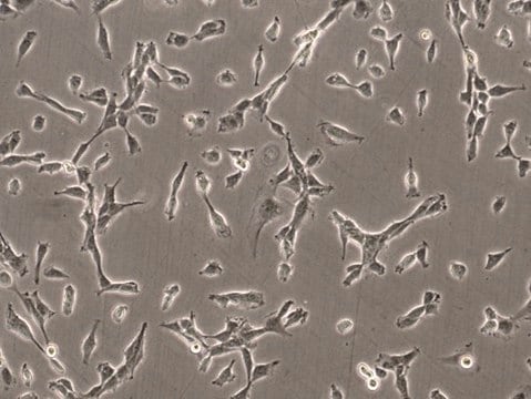
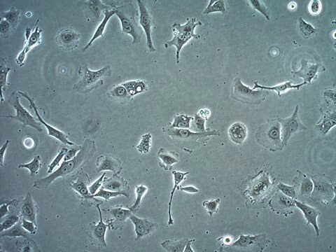
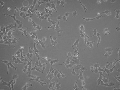
All Photos(3)

Key Documents

SCC201

Sigma-Aldrich

LOX-IMVI Human Melanoma Cell Line

Human

Synonym(s):

LOX/IMVI, LOX IMVI, LOXIM-VI, LOXIMVI

Sign Into View Organizational & Contract Pricing


About This Item

UNSPSC Code:
41106514
eCl@ss:
32011203
NACRES:
NA.81

product name

LOX-IMVI Human Melanoma Cell Line, LOX-IMVI human melanoma cell line is an excellent model for probing mechanisms of metastasis and for evaluation of chemotherapies.

biological source

human

technique(s)

cell culture | mammalian: suitable

General description

Malignant melanoma is highly treatable when diagnosed early, but can spread rapidly if undetected, resulting in the highest mortality among skin cancer types. Lymph nodes and lungs are the most common sites of melanoma metastases , and because of the poor prognosis and limited efficacy of current treatments, metastatic melanoma has generated intense interest as a target for therapeutic intervention.
The LOX-IMVI human melanoma cell line is widely used as an in vitro model system to study tumor metastasis and to test for chemosensitivity to potential anti-cancer compounds. LOX-IMVI cells exhibit a tendency to form lung metastases in nude mice independent of the inoculation site , and mortality of experimental animals is observed within 3-5 weeks of LOX-IMVI cell injection . LOX-IMVI cells are genetically characterized by lack of the Y chromosome and trisomy 7 , and are heterozygous for the BRAF V600E melanoma driver mutation . LOX-IMVI cells are amelanotic and express the human melanoma marker GD3 ganglioside, a factor in metastatic potential of malignant melanoma .

Source:
The LOX-IMVI human melanoma cell line was established from a subcutaneous xenograft in nude mice from a lymph node metastasis of a 58-year-old Caucasian male patient with malignant amelanotic melanoma .

Cell Line Description

Cancer Cells

Application

LOX-IMVI human melanoma cell line is an excellent model for probing mechanisms of metastasis and for evaluation of chemotherapies.
Research Category
Cancer
This product is intended for sale and sold solely to academic institutions for internal academic research use per the terms of the “Academic Use Agreement” as detailed in the product documentation. For information regarding any other use, please contact licensing@emdmillipore.com.

Quality

• Each vial contains ≥ 1X10⁶ viable cells.
• Cells are tested negative for HPV-16, HPV-18, Hepatitis A, B, C, and HIV-1 & 2 viruses by PCR.
• Cells are verified to be of human origin and negative for inter-species contamination from rat, mouse, chinese hamster, Golden Syrian hamster, and non-human primate (NHP) as assessed by a Contamination CLEAR panel by Charles River Animal Diagnostic Services.
• Cells are negative for mycoplasma contamination.
• Each lot of cells is genotyped by STR analysis to verify the unique identity of the cell line.

Storage and Stability

Store in liquid nitrogen. The cells can be cultured for at least 10 passages after initial thawing without significantly affecting the cell marker expression and functionality.

Disclaimer

Unless otherwise stated in our catalog or other company documentation accompanying the product(s), our products are intended for research use only and are not to be used for any other purpose, which includes but is not limited to, unauthorized commercial uses, in vitro diagnostic uses, ex vivo or in vivo therapeutic uses or any type of consumption or application to humans or animals.

Storage Class

10 - Combustible liquids

wgk_germany

WGK 1

flash_point_f

Not applicable

flash_point_c

Not applicable


Certificates of Analysis (COA)

Search for Certificates of Analysis (COA) by entering the products Lot/Batch Number. Lot and Batch Numbers can be found on a product’s label following the words ‘Lot’ or ‘Batch’.

Already Own This Product?

Find documentation for the products that you have recently purchased in the Document Library.

Visit the Document Library

AP-1 transcription factor network explains diverse patterns of cellular plasticity in melanoma cells.
Comandante-Lou, et al.
Cell Reports, 40, 111147-111147 (2022)
Kazunori Hamamura et al.
Proceedings of the National Academy of Sciences of the United States of America, 102(31), 11041-11046 (2005-07-26)
Although ganglioside GD3 levels are highly elevated in malignant melanomas, the role of GD3 in melanomas' malignant properties has not been clearly shown. To investigate this problem, we genetically generated GD3-positive (GD3+) transfectant cells from a GD3-negative (GD3-) mutant line
I Kjønniksen et al.
Cancer research, 49(18), 5148-5152 (1989-09-15)
Experimental lung metastases regularly developed in athymic Han:rnu/rnu Rowett rats after i.v. injection of LOX human malignant melanoma cells. When 5 x 10(5) tumor cells were injected into 4-week-old rats, 89% of the animals died of lung tumors, with a
Jana Jandova et al.
Molecular carcinogenesis, 61(6), 603-614 (2022-04-14)
Molecularly targeted therapeutics have revolutionized the treatment of BRAFV600E -driven malignant melanoma, but the rapid development of resistance to BRAF kinase inhibitors (BRAFi) presents a significant obstacle. The use of clinical antimalarials for the investigational treatment of malignant melanoma has
James J Miller et al.
Scientific reports, 11(1), 8213-8213 (2021-04-17)
Plasmin is the key enzyme in fibrinolysis. Upon interaction with plasminogen activators, the zymogen plasminogen is converted to active plasmin. Some studies indicate plasminogen activation is regulated by cation-independent mannose 6-phosphate receptor (CI-MPR), a protein that facilitates lysosomal enzyme trafficking

Our team of scientists has experience in all areas of research including Life Science, Material Science, Chemical Synthesis, Chromatography, Analytical and many others.

Contact Technical Service