
추천 제품


생물학적 소스
mouse
Quality Level
결합
peroxidase conjugate
항체 형태
purified immunoglobulin
항체 생산 유형
primary antibodies
클론
BMG-his-1, monoclonal
양식
lyophilized
포장
pkg of 50 U
제조업체/상표
Roche
동형
IgG1
저장 온도
2-8°C
관련 카테고리
일반 설명
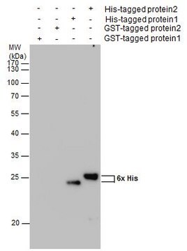
Anti-His6-Peroxidase is a monoclonal antibody to His6-tagged proteins, conjugated to horseradish peroxidase. Anti-His6-Peroxidase specifically recognizes an epitope of six consecutive histidine residues of both natural and recombinant proteins. The antibody reacts with native and denatured histidine-tagged fusion proteins independent of the epitope-sequence location; however, it preferentially recognizes the C-terminal His6 epitope with high sensitivity.
특이성
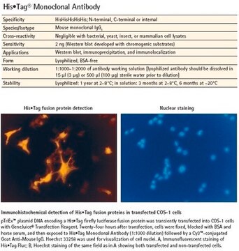
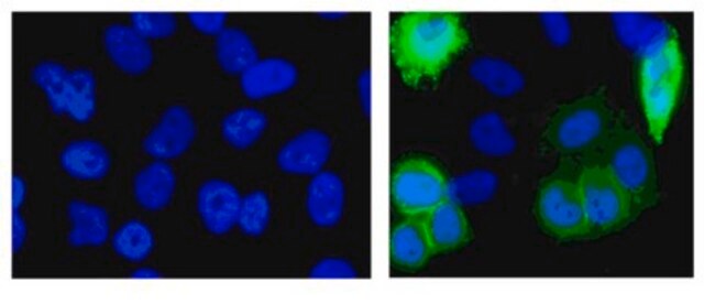
Anti-His6-Peroxidase specifically recognizes an epitope of six consecutive histidine residues (His6) in natural and recombinant proteins. It reacts with both native and denatured histidine-tagged fusion proteins, independent of location of the epitopesequence. However, Anti-His6-Peroxidase preferentially recognizes the C-terminal His6 epitope with high sensitivity. Anti-His6 allows specific and sensitive detection of histidine-tagged proteins irrespective of the expression system used.
The antibody recognizes an epitope of six consecutive histidine residues (His6) in natural and recombinant proteins. It reacts with both native and denatured histidine-tagged fusion proteins, independent of the epitope-sequence location.
애플리케이션
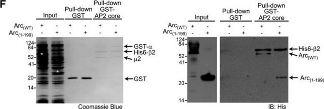
Anti-His6-Peroxidase has been used in in vitro serum stability of fusion toxins, pull-down assay and western blotting.
Anti-His6-Peroxidase is used for the detection of His6-tagged proteins in:
- ELISA (enzyme-linked immunosorbent assay)
- Western blot
품질
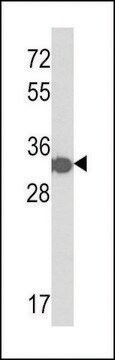
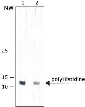
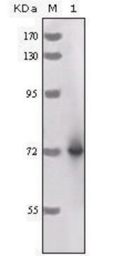
Function test: Western blot using extracts from cell line expressing a recombinant His6-tagged protein.
제조 메모
Working concentration: Working concentration of conjugate depends on application and substrate.
The following concentrations should be taken as a guideline:
The following concentrations should be taken as a guideline:
- ELISA: 100 mU/ml
- Western blot: 100mU/ml
재구성
Add 1 ml double-distilled water to a final concentration of 50 U/ml.
Reconstitution should be performed for at least 10 minutes.
Reconstitution should be performed for at least 10 minutes.
기타 정보
For life science research only. Not for use in diagnostic procedures.
적합한 제품을 찾을 수 없으신가요?
당사의 제품 선택기 도구.을(를) 시도해 보세요.

신호어
Warning
유해 및 위험 성명서
Hazard Classifications
Skin Sens. 1
Storage Class Code
11 - Combustible Solids
WGK
WGK 1
Flash Point (°F)
does not flash
Flash Point (°C)
does not flash
이미 열람한 고객
Hiroko Ideo et al.
The Journal of biological chemistry, 284(39), 26493-26501 (2009-07-29)
Galectins are a family of beta-galactoside-binding proteins that are widely found among animal species and that regulate diverse biological phenomena. To study the biological function of glycolipid-binding galectins, we purified recombinant Caenorhabditis elegans galectins (LEC-1-11) and studied their binding to
Andra-Octavia Roman et al.
Nature communications, 13(1), 876-876 (2022-02-17)
The membrane receptor kinases HAESA and HSL2 recognize a family of IDA/IDL signaling peptides to control cell separation processes in different plant organs. The homologous HSL1 has been reported to regulate epidermal cell patterning by interacting with a different class
Elaheh Gheybi et al.
Cancer reports (Hoboken, N.J.), 6(3), e1745-e1745 (2022-10-28)
CD44, as a tumor-associated marker, can be used to detect stem cells in breast cancer. While CD44 is expressed in normal epithelial cells, carcinoma cells overexpress CD44. In the current study, we designed a recombinant protein that included the variable
KaiC intersubunit communication facilitates robustness of circadian rhythms in cyanobacteria
Kitayama Y, et al.
Nature Communications, 4 (2013)
Clare Sheridan et al.
Molecular cell, 31(4), 570-585 (2008-08-30)
Bax and Bak promote apoptosis by perturbing the permeability of the mitochondrial outer membrane and facilitating the release of cytochrome c by a mechanism that is still poorly defined. During apoptosis, Bax and Bak also promote fragmentation of the mitochondrial
자사의 과학자팀은 생명 과학, 재료 과학, 화학 합성, 크로마토그래피, 분석 및 기타 많은 영역을 포함한 모든 과학 분야에 경험이 있습니다..
고객지원팀으로 연락바랍니다.